Hemangioma on dog
Hemangioma On Dog. Unfortunately hemangiosarcoma symptoms often dont become obvious until the cancer has. What is Hemangioma in Dogs. Several var-iants of hemangiomas are described such as capillary and cavernous hemangiomas infiltrative hemangioma arteriovenous hemangioma gran-ulation tissuetype hemangioma spindle cell hem-. We were unable to load Disqus Recommendations.
Jebas Org From
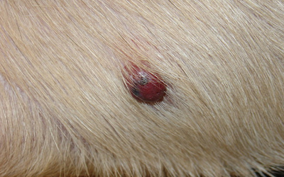
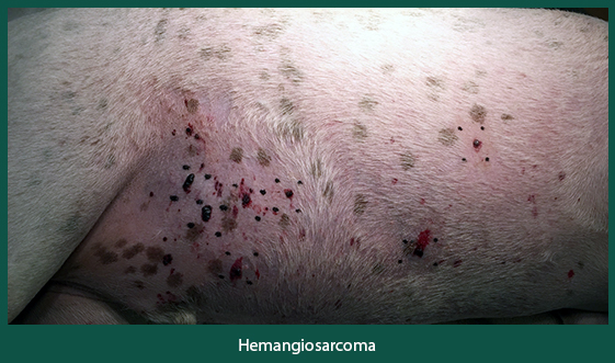
Youll most likely find a hemangioma on the dogs trunk or legs especially hairless areas like the lower abdomen. In vertebral hemangioma it has been reported to demonstrate from ametabolic cold vertebra to low 18 F-FDG uptake on PETCT images. Hemangiomas are the most common benign solid mass in the liver and are often incidentally discovered during imaging. Hemangiomas are lesions of the vascular system that are formed by the cells that are responsible for forming blood vessels or endothelial cells. What is Hemangioma in Dogs. Staging searching for potential spread to other locations in the body should be pursued with any diagnosis of hemangiosarcoma.
Although plain radiography CT MRI and angiography may provide anatomic extent and be pathognomonic FDG-PET and FMT-PET may be the most reliable among the studied imaging modalities for differentiating benign hemangiomas from other soft tissue tumors especially malignant neoplasms.
Hemangiomas in dogs are generally benign soft tissues and skin tumors while sarcomas are malignant tumors developing in the soft tissues. Up to 70 of dogs with splenic tumors have hemangiosarcoma. A hemangioma is a benign mass in the spleen of canines. Ray- Hemangiomas are a benign lump formed by blood vessel tissues that occur in the skin. Spontaneous tumors of blood vessel endothelial cells have been described commonly in the dog less frequently in the cat and horse and sporadically in most other domestic species. Hemangiomas are lesions of the vascular system that are formed by the cells that are responsible for forming blood vessels or endothelial cells.
 Source: topdogtips.com
Hemangiomas in dogs are generally benign soft tissues and skin tumors while sarcomas are malignant tumors developing in the soft tissues. Hemangiomas are growths that are made up of many tiny blood vessels bunched together. These growths usually dont appear until at least middle age. This type of growth a cutaneous hemangioma is a neoplasm on the skin that is benign. Histologically endothelial tumors in animals are most commonly designated as angiomatosis he-mangiomas and hemangiosarcomas.
 Source: cliniciansbrief.com
Hemangioma has been known to be a metabolically stable benign lesion on PET. However rarely they may have atypical imaging features and appear hypermetabolic. In this dog synovial hemangioma evident as a soft tissue mass on radiographs was associated with chronic lameness and hemarthrosis and resolved. Several var-iants of hemangiomas are described such as capillary and cavernous hemangiomas infiltrative hemangioma arteriovenous hemangioma gran-ulation tissuetype hemangioma spindle cell hem-. Ray- Hemangiomas are a benign lump formed by blood vessel tissues that occur in the skin.
 Source: topdogtips.com
Hemangiomas are a type of benign tumor of the blood vessels or skin. Skeletal hemangioma is one of the common benign conditions which are typically ametabolic on FDG PET CT with no uptake on bone scan. Skin Cancer Hemangiosarcoma in Dogs PetM. Hemangioma has been known to be a metabolically stable benign lesion on PET. Veterinary Surgery 38463466 2009 BRIEF COMMUNICATION Synovial Hemangioma in a Dog J.
 Source: topdogtips.com
6 In the dog hemangiomas are typically benign solitary deep dermal tumors whereas hemangiosarcomas often present as a disseminated malignancy involving the spleen. This type of growth a cutaneous hemangioma is a neoplasm on the skin that is benign. Histologically endothelial tumors in animals are most commonly designated as angiomatosis he-mangiomas and hemangiosarcomas. They are more common in dogs but do occur in cats as well. This birthmark is usually just a small mark on the face trunk or extremities arms and legs.
 Source: researchgate.net
Hemangiomas consist of benign blood-filled vascular spaces lined with epithelia and containing fibrous septa. Hemangiomas are lesions of the vascular system that are formed by the cells that are responsible for forming blood vessels or endothelial cells. Additionally since it is full of blood vessels it may bleed excessively if accidentally injured. Unfortunately hemangiosarcoma symptoms often dont become obvious until the cancer has. Up to 70 of dogs with splenic tumors have hemangiosarcoma.
 Source: vcahospitals.com
It resembles a blood blister or angiokeratoma. Several var-iants of hemangiomas are described such as capillary and cavernous hemangiomas infiltrative hemangioma arteriovenous hemangioma gran-ulation tissuetype hemangioma spindle cell hem-. The spleen is located below your dogs stomach and is in the shape of a tongue. Hemangiomas are lesions of the vascular system that are formed by the cells that are responsible for forming blood vessels or endothelial cells. This forms a mass in your dogs tummy and causes pain.
Source:
They are more common in dogs but do occur in cats as well. They are more common in dogs but do occur in cats as well. The spleen is located below your dogs stomach and is in the shape of a tongue. Red blood cells are mass-produced and do not expel through the spleen as they should. It originates either in the dermis or the subcutaneous layer of the dogs skin.
 Source: dermvets.com
Ray- Hemangiomas are a benign lump formed by blood vessel tissues that occur in the skin. Although plain radiography CT MRI and angiography may provide anatomic extent and be pathognomonic FDG-PET and FMT-PET may be the most reliable among the studied imaging modalities for differentiating benign hemangiomas from other soft tissue tumors especially malignant neoplasms. Spontaneous tumors of blood vessel endothelial cells have been described commonly in the dog less frequently in the cat and horse and sporadically in most other domestic species. We were unable to load Disqus Recommendations. IGNACIO ARIAS DVM CRISTIAN TORRES DVM and DANIEL SAEZ DVM ObjectiveTo report arthroscopic diagnosis and treatment of synovial hemangioma in a dog.
 Source: topdogtips.com
Hemangiomas are lesions of the vascular system that are formed by the cells that are responsible for forming blood vessels or endothelial cells. Skeletal hemangioma is one of the common benign conditions which are typically ametabolic on FDG PET CT with no uptake on bone scan. It resembles a blood blister or angiokeratoma. 6 In the dog hemangiomas are typically benign solitary deep dermal tumors whereas hemangiosarcomas often present as a disseminated malignancy involving the spleen. Staging searching for potential spread to other locations in the body should be pursued with any diagnosis of hemangiosarcoma.
 Source: researchgate.net
Red blood cells are mass-produced and do not expel through the spleen as they should. Additionally since it is full of blood vessels it may bleed excessively if accidentally injured. Skeletal hemangioma is one of the common benign conditions which are typically ametabolic on FDG PET CT with no uptake on bone scan. It originates either in the dermis or the subcutaneous layer of the dogs skin. AnimalStandard Poodle 8-year-old neutered male.
 Source: whole-dog-journal.com
As alluded to above three retrospective studies found that dogs with non-traumatic hemoperitoneum 15 and with hemoperitoneum due specifically to splenic masses 3 had a greater prevalence of hemangiosarcoma 633-704 when compared to dogs in large-scale retrospective studies which included all splenic samples whether associated with. Skin Cancer Hemangiosarcoma in Dogs PetM. We were unable to load Disqus Recommendations. Some hemangiomas are more serious. Hemangiosarcoma or cancer of the blood vessels is a type of tumor occurring more often in canines than any other animal.
 Source: merckvetmanual.com
Hemangiomas are growths that are made up of many tiny blood vessels bunched together. What is Hemangioma in Dogs. Hemangioma has been known to be a metabolically stable benign lesion on PET. Hemangiomas are the most common benign solid mass in the liver and are often incidentally discovered during imaging. Ulaner MD PhD FACNM in Fundamentals of Oncologic PETCT 2019.
If you find this site helpful, please support us by sharing this posts to your own social media accounts like Facebook, Instagram and so on or you can also bookmark this blog page with the title hemangioma on dog by using Ctrl + D for devices a laptop with a Windows operating system or Command + D for laptops with an Apple operating system. If you use a smartphone, you can also use the drawer menu of the browser you are using. Whether it’s a Windows, Mac, iOS or Android operating system, you will still be able to bookmark this website.


