Vein on dogs front leg
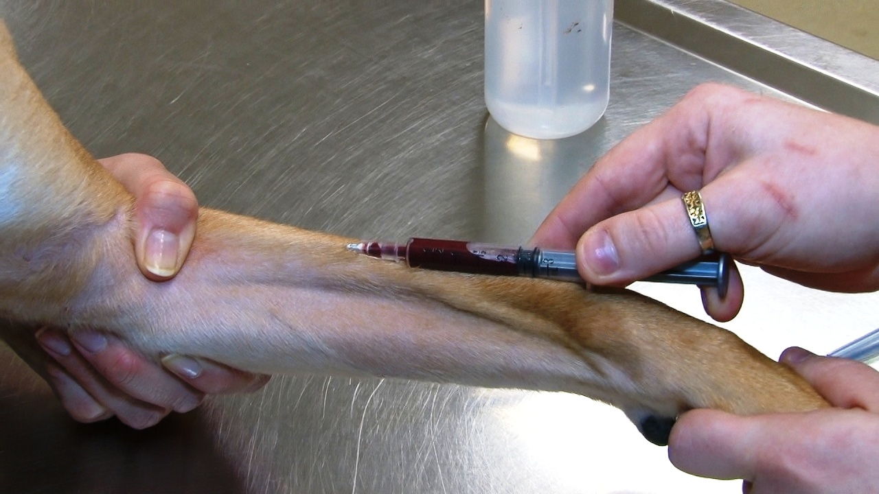
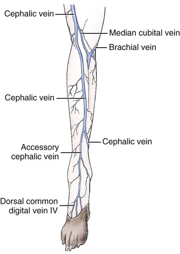
Vein On Dogs Front Leg. The image below shows the cephalic vein. No more than eight blood samples four from either leg should be taken in any 24 hour period. That way they can determine if treatment is necessary. A 22-gauge needle attached to a 3-ml syringe is slowly inserted at a 25-degree angle through the skin and into the vein.
 A Photograph Showing Distribution Of Arteries Veins And Nerves On Download Scientific Diagram From researchgate.net
It is reinforced by strong fascia. Medial scapula and scapular cartilage Action. Surgery might be an option and there are certain drugs that might be applied. Ad Laser removal without surgery. Occlude the cephalic vein by having your helper hold off the vein. The venipuncture should be performed in the middle 13 of the jugular vein with a needle and syringe.
Phlebitis is characterized by a condition known as superficial thrombophlebitis which refers to an inflammation of superficial veins or veins close to the surface of the body.
Forepaw of a front dog leg. Phlebitis is generally due to an infection or because of thrombosis – the formation of a clot or thrombus inside a blood vessel which in turn. The cephalic vein is located on the front of the foreleg the dorsal surface. In this case I have put a tourniquet on the elbow so that the vein fills with. Once the patient is restrained the phlebotomist grabs the leg places a thumb lateral to the vein and pulls the skin distally to stabilize the vein. Surgery might be an option and there are certain drugs that might be applied.
 Source: quizlet.com
While dogs can develop cancerous tumors if you find a growth on your dogs skin many are treatable. The venipuncture should be performed in the middle 13 of the jugular vein with a needle and syringe. The veterinarian will first need to perform a physical examination to try to determine the type and extent of the injury. Two people are required to take the blood sample. Only one third is.
 Source: chegg.com
The tumors may be either benign or deadly and according to the diagnosis a treatment may be established. The veterinarian will first need to perform a physical examination to try to determine the type and extent of the injury. It is reinforced by strong fascia. The dogs leg is being held. The image below shows the cephalic vein.
 Source: beattiepethospitalhamilton.com
Phlebitis is characterized by a condition known as superficial thrombophlebitis which refers to an inflammation of superficial veins or veins close to the surface of the body. A 22-gauge needle attached to a 3-ml syringe is slowly inserted at a 25-degree angle through the skin and into the vein. The dog is restrained close to the body of the holder. The wrist is also called the carpus. You will find seven bones in the carpus of a dogs front leg that arranges in two rows.
 Source: researchgate.net
Supporting the weight of the trunk. Once the patient is restrained the phlebotomist grabs the leg places a thumb lateral to the vein and pulls the skin distally to stabilize the vein. On the subject of leg muscles lets have a look at the leg as a whole in a little more detail. Cost of Front Leg Injury in Dogs. C4 to 10th rib Insertion.
 Source: atdove.org
While dogs can develop cancerous tumors if you find a growth on your dogs skin many are treatable. This veterinary anatomical atlas includes selected labeling structures to help student to understand and discover animal anatomy skeleton bones muscles joints viscera respiratory system. The vein runs under the skin between the carpal wrist joint and the elbow. 21G or wider needle. Ad Laser removal without surgery.
 Source: vetstore.in
The cervical portion can retract the limb and the caudal portion can advance the limb. Dog anatomy comprises the anatomical studies of the visible parts of the body of a domestic dogDetails of structures vary tremendously from breed to breed more than in any other animal species wild or domesticated as dogs are highly variable in height and weight. Two thirds of a dogs body weight is carried on their front legs. Our dog had his blood drawn on Tuesday August 20 5 days ago. The omobrachial vein v.
 Source: youtube.com
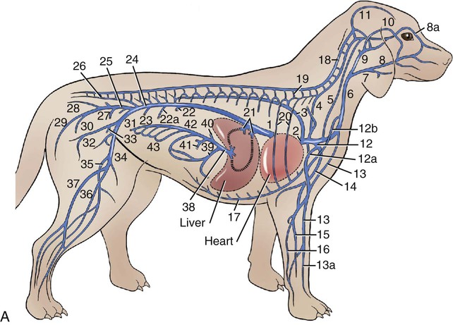
Brachiocephalicus and enters the lateral surface of the. Omobrachialis formerly called the proximal communicating vein of the cephalic vein leaves the axillobrachial vein approximately 2 cm proximal to the cephalic vein and runs superficially at first on the m. Another name of the dog forepaw is maneus. The omobrachial vein v. It then arches cranially and medially crosses the m.
 Source: canstockphoto.com
The smallest known adult dog was a Yorkshire Terrier that stood only 63 cm 25 in at the shoulder 95 cm. The cervical portion can retract the limb and the caudal portion can advance the limb. Ad Laser removal without surgery. Anatomy of the dog - Illustrated atlas This modules of vet-Anatomy provides a basic foundation in animal anatomy for students of veterinary medicine. The omobrachial vein v.

This is done by placing your hand under the elbow so that the dog cannot move its leg back and taking your thumb over the top of the leg and applying pressure so that the cephalic. It draws the trunk forward when the limb is fixed. The cervical portion can retract the limb and the caudal portion can advance the limb. Take 2-3 fingers and place them longways along the top of the forearm to feel the vein and trace how it is going down the leg. The cephalic vein is wiped with alcohol.

Our dog had his blood drawn on Tuesday August 20 5 days ago. One for restraining and raising the vein of the dog and one for taking the blood sample. Phlebitis is generally due to an infection or because of thrombosis – the formation of a clot or thrombus inside a blood vessel which in turn. C4 to 10th rib Insertion. Supporting the weight of the trunk.
 Source: veteriankey.com
The front legs and neck up to the point of the mandible should be in a vertical plane. By renowned surgeon Dr. If you cant tell if it is the vein have your holder release their grip. The venipuncture should be performed in the middle 13 of the jugular vein with a needle and syringe. While dogs technically do not have arms they do have elbows and wrists.
 Source: veteriankey.com
This is done by placing your hand under the elbow so that the dog cannot move its leg back and taking your thumb over the top of the leg and applying pressure so that the cephalic. The muzzle is held away from the face of the holder and the person placing the catheter. Two people are required to take the blood sample. The vet tried the left front leg first and our dog shrieked immediately not his usual reaction. Branch of brachial plexus Origin.
If you find this site adventageous, please support us by sharing this posts to your own social media accounts like Facebook, Instagram and so on or you can also save this blog page with the title vein on dogs front leg by using Ctrl + D for devices a laptop with a Windows operating system or Command + D for laptops with an Apple operating system. If you use a smartphone, you can also use the drawer menu of the browser you are using. Whether it’s a Windows, Mac, iOS or Android operating system, you will still be able to bookmark this website.

